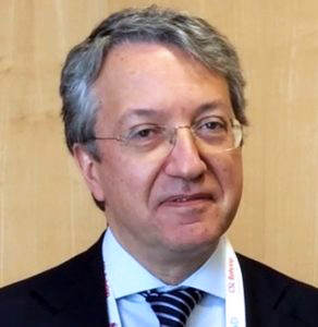
By: Dr. Robert Bard
Cancer Imaging Specialist for the NY Cancer Resource Alliance Part I
The record of cancer treatment advancements carry a significant debt to a community of Italian clinical pioneers recognized for their extensive contribution to the screening, imaging and diagnostic innovations. Names such as Drs. Luigi Solbiati, Carlo Martinoli and Rodolfo Campani are some of the top names who helped pave the movement for a much improved detection of cancer tumors and other subdermal disorders.
The Birth of Medical Radiology
Since the discovery of the X-Ray in 1890, scientists worldwide found the drive to mobilize diagnostic science into a non-invasive direction. Scanning technology carried the potential to save lives by enabling images of physiological issues underneath the skin non-invasively or without any cutting. But it wasn’t until 1977 that the results of radiological imaging advanced to the capacity of cancer detection as the Magnetic Resonance Imaging (MRI) was developed by Dr. Raymond Damadian, who performed the first full body scan to diagnose cancer and refining the focus of modern radiology.
The Bracco Imaging Group, headquartered in Milan, Italy, established the first multinational healthcare group in 1927 and heavily supported many contributions to clinical diagnostic science including the launch of the advancement of contrast agents for all imaging solutions. It is this material injected in the bloodstream that allowed a significant improvement in identifying tumor cells.
One of the leading pioneers in this study was Professor Rodolfo Campani who started the first world study of ultrasound contrast agents in humans in Italy in 1990. The Scientific Journal of the Italian Society of Radiology “La Radiologia Medica” (reported in May 1993) presented the results of the five Italian experimentation centers under his coordination at the Institute of Radiology in the University of Pavia in Italy. He was credited for developing the first (non-radiation based) ultrasound contrast agent which are used to show blood flow in tumors, elevating the power of the ultrasound to out-perform MRIs, X-rays and CT Scans. Implementation of this contrast was a generational leap in advantages because it made the tumor vascularity much more easily visible using the injectable and safe ultrasound contrast agent. Unlike the MRI contrasts, which contained heavy metals, the ultrasound contrast was comprised of air bubbles and microalbumin. In a 1994 journal, this contrast was first used in liver cancers where vascularity is highlighted inside the liver. With this same time span, Carlo Martinoli, MD, of Genoa, arose as another contributor to the widespread use of ultrasound technology by emphasizing the effects of ultrasound imaging of the musculoskeletal and peripheral nervous systems. He helped shape modern medical education to recognize and include the studies of ultrasound and musculoskeletal radiology and co-authored vital textbooks on the subject which are still in use today in over 22 countries.
Italian History of Tracking Blood Flow
Leonardo da Vinci detailed over 500 years ago about the way the blood flowed in and out of the heart and showed how the valves worked. It wasn’t until 1960 that the medical community caught up to da Vinci and confirmed all this time that he was remarkably correct! A jump to the 1990s became a pivotal period as European imaging has standardized the non-invasive ultrasound technology to be a major screening protocol for cancer investigation.
Professor Luigi Solbiati of Milan, specialized in the study of blood flow of cancers and published his discoveries about the major differences between malignant and benign tumor vessels. This study helped to shape the way noninvasive imaging protocols like the newer Doppler sonography and MRI diagnostic techniques identify cancers. He pioneered ultrasound-guided aspiration biopsies (1979), ethanol injection (1983), radiofrequency ablation with cool-tip electrodes (1995) and microwaves (2009) of solid tumors and fusion imaging for the guidance of interventional procedures (2003).
Modern studies confirm that metastasis (the spread of cancer to other areas of the body) and cell migration is mobilized by blood flow and it is here that diagnostic protocols examine other clues as to the condition of the existing tumors. Digital ultrasound imaging with blood flow technology, the Doppler blood flow and the contrast enhanced blood flow was pioneered and developed by Rodolfo Campani and Luigi Solbiati at the University of Pavia Medical Center in Italy, where they introduced worldwide, the performance of the Doppler Ultrasound as a viable and more accurate technology for diagnosing tumors and assessing tumor response in many different areas.


